 |
|
1 Comment
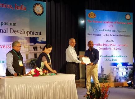
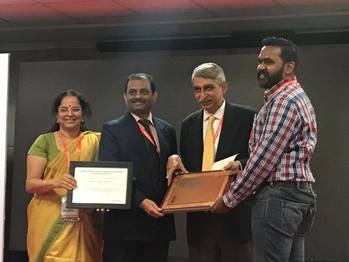 Dr. Vijay Harish Somasundaram, Clinical Asst. Professor in department of Nuclear Medicine & PET CT who has completed PhD under the guidance of Dr. Manzoor K (ACNS) & Dr. P Shanmugasundaram (AIMS); presented a part of his doctoral work at the 15th annual ANMPI (Association of Nuclear Medicine Physicians of India) conference held in association with the ICC (Indian Cancer Congress) 2017 conference at Bangalore (8th-12th Nov 2017).His paper titled: "Novel, biodegradable, radiopaque microbeads for Transarterial Radioembolization of liver tumors" was awarded to prestigious 'Dr. (Maj Gen) Lakshmipathy Award' for best oral presentation of original research.His work was also selected as one of the best three works from among 46 entries from all over India, for a 'Pitch - Fest' of innovative ideas that can be potentially translated for clinical use.This competition was judged by some of the top venture capitalists of the country and Dr. Vijay Harish won the second prize of cash award one Lakh rupees and 1 year mentorship to translate this work. 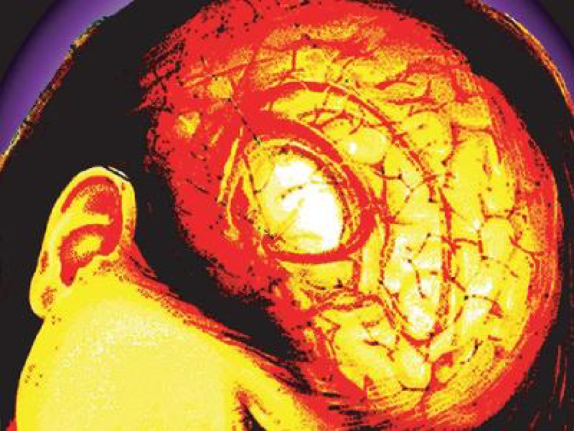 TNN | Mar 31, 2017, 12.27 PM IST
COIMBATORE: Kochi-based scientists have now developed a small wafer made of nanofibers that delivers chemotherapy and dissolves cancerous brain tumours. Made in India, the product that will cost just Rs 50,000, one sixth of the price of imported devices, is expected to hit the market in five years, the scientists said. The implant was developed by a group from Amrita Institute of Nanomedicine with Rs 5 crore funding by the department of science and technology, government of India. The research was published recently in scientific journal, Nature. Oncologists say there is risk of reoccurrence of high grade brain tumours, despite surgery, chemotherapy drugs and radiation. "Cells that are invisible to the surgeon will remain in the tumour resected margin. Some of these cells may be resistant to chemotherapy and radiation. These cells multiply and cancer recurs. In most patients recurrence happens within the first two years," said Manzoor Koyakutty, a professor at Amrita Institute of Nanomedicine, who developed the implant. The nano-wafer which will be placed in the tumour cavity has the potential to destroy residual tumour cells. It will also release chemotherapy drugs slowly over two month and fade away, said Koyakutty. "The drug it elutes will significantly inhibit tumour recurrence and extend the life of the patient," he said. What has excited oncologists and neurosurgeons even more about the flexible and biocompatible wafer is its cost. Sri Ramachandra Medical University consultant neurosurgeon Dr K Selva Kumar said that similar implants are available abroad. "They are generally purchased from Germany, Japan and the USA for Rs 3-4 lakh but their effect lasts for about four years," he said. Koyakutty said it was a challenge to get the wafer to release the drug in a continuous and controlled manner. "Products in the US and European market deliver drugs in the brain for just upto a week, but we wanted our implant to deliver the drug up to two months at a lower cost," Koyakutty said. Scientists say indigenous wafer has undergone extensive testing in animal models and no adverse effects were seen."We are now testing the drug on larger animals such as pigs," said Koyakutty. If these trials succeed, the wafer will have to undergo clinical trials before it hits the market. Glioma -a tumour that starts in the brain or spine -accounts for 5-6% of all tumours in India. The disease has high mortality rates because oral chemotherapy does not effectively kill tumours cells in the brain. "Only 30-32% of the drug reaches the brain. But a nano-implant can elute 100% drug directly in to the tumour," said Dr Dilip Panikar, head of neuro surgery at Amrita Hospital. http://timesofindia.indiatimes.com/city/coimbatore/nano-wafers-to-prevent-brain-tumour-recurrence/articleshow/57935649.cms  New drug delivery process gives hope to leukemia patientsTNN | Oct 3, 2016, 03.45 AM IST Coimbatore: It's common to see leukemia patients develop resistance towards the drug during treatment, leaving them with bone marrow transplant as the only choice of cure. Bone marrow transplant, however, is not an easy option in-part due to difficulties in getting a matching donor, doctors say. Manzoor Koyakutty along with Shantikumar Nair and Dr Pavithran K, researchers at Amrita University's Centre for Nanoscienes and Nanomedicine, have identified a new drug delivery system that they say could eliminate the drug resistance in leukemia. Chronic myeloid Leukemia is a condition where the body produces excess white blood cells. These cells accumulate around the bone marrow and interfere with the production of blood cells. According to a study, only 5% patients with adult leukemia in India survive after diagnosis. There are drugs available in the market to treat leukemia. "If the disease progresses while on any of these drugs, then the only option currently available to them is bone marrow transplant or supportive care," said the head of medical oncology at Amrita Institute of Medical Sciences, Kochi, Dr K Pavithran. A patient develops resistance towards a drug due to gene mutation when the cancer cells proliferate. So a more efficient drug can eliminate resistance developing, the researchers say. The scientists along with their PhD scholars worked on a core-shell nanomedicine technique to improve the action of the drug. In this technique, the scientists have used two components - the drug and an inhibitor. "The nanoparticle is divided into two compartments - shell and core. The drug is embedded in the shell, and the inhibitor is embedded in the core. When the nanoparticle comes in contact with cancer cells, it dissolves and the drug begins action," said professor Shantikumar Nair. "The inhibitor prevents the cancer cells from evading the drug's action. This improves the efficiency of the drug," he added. The two drugs are not given together in free form since they can react with each other and create toxicity and increase the chances of reaction with blood components. "If the two drugs are given separately they may not be effective," said Nair. The technology was awarded a US patent in July 2016. At present, the technology has been tested with human samples, and the four-year-long research work was carried out with the help of Rs5crore granted by the department of biotechnology (DBT). "In addition, we are likely to receive Rs7crore from DBT to carry out studies on animals and perform human trials. Amrita will also invest Rs3crore to set up a good manufacturing process (GMP) and a clean room facility," said Nair, adding that it will take another five years for the scientists to come out with the drug. http://timesofindia.indiatimes.com/city/coimbatore/New-drug-delivery-process-gives-hope-to-leukemia-patients/articleshow/54647331.cms
 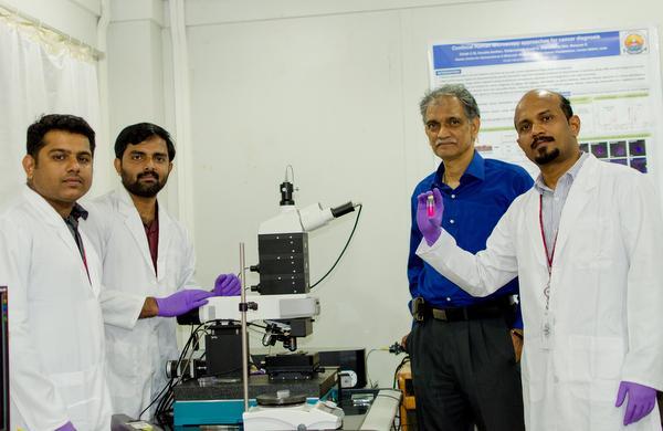 (From left) Ranjith Ramachandran, Girish C.M, Dr. Shantikumar Nair and Dr. Manzoor Koyakutty have produced nanoparticles containing the drug that can absorb three times more light in the near IR.
The prolonged period of stability of the drug provides a longer window for therapy. Scientists at the Amrita Centre for Nanosciences and Molecular Medicine at Amrita University, Kochi, have come a step closer to using photodynamic therapy for treating residual cancer cells of a high-grade brain tumour (glioblastoma). Photodynamic therapy uses a photosensitive drug that becomes active under the action of light and converts molecular oxygen into reactive oxygen species that kill cancer cells. While the photosensitive drug injected into the body intravenously is not cancer-cell specific and is less efficient in absorbing light to generate reactive oxygen species, scientists at the Amrita Institute have turned to nanotechnology and used light in the near-infrared region to achieve better results. Light in the near-infrared region can penetrate to about 0.8 cm into body tissues. “The drug encapsulated in a nanoparticle has peptides functionalised on its surface and is selectively absorbed only by cancer cells. The nanoparticles containing the drug have better ability to kill cancer cells as they absorb three times more light in the near IR region than the free drug,” says Dr. Manzoor Koyakutty from the Amrita Centre for Nanosciences and Molecular Medicine. “We have incorporated a photosensitiser into the nanoparticle to enhance the light absorption capacity.” Dr. Koyakutty had carried out studies on mice while he was in Erasmus University Rotterdam, the Netherlands. A unique chemical bonding used for anchoring the photosensitiser to the nanoparticle increases the stability of the drug by as much as nine times, says Dr. Koyakutty. The prolonged period of stability of the drug provides a longer window for therapy; light in the near-infrared range can now be given in fractions to activate the drug at regular intervals. “Even if the tumour mass is removed from the brain, some residual cancer cells will be present near the tumour site. We can’t remove healthy brain tissue that contains some cancer cells. So once you remove the tumour mass we can apply the nanoparticles containing the drug at the site where the tumour was present and use near-IR [light] to kill cancer cells in the neighbouring areas,” says Dr. Shantikumar Nair, Director of the Centre. “Recurrence is very high in the case of glioma. Patients don’t have a cure if it recurs. So if recurrence can be prevented patients can have substantial additional life,” says Dr. Nair. “Photodynamic therapy is a well known treatment option but can be used only when cancers are near the skin as light cannot penetrate deep. So it has not become a popular treatment option but has potential,” says Dr. Rajiv Sarin, Professor, Radiation Oncology and in-charge of Cancer Genetics Unit at Tata Memorial Hospital, Mumbai. “Getting clinical benefits in patients with cancer is more challenging as cancer is seldom near the surface.” The scientists are planning to undertake studies on mice and then larger animals.  Coimbatore: Four years ago, scientists Shantikumar V Nairand Manzoor Koyakutty ofAmrita University's Kochi-based Centre for nanomedicine were using lasers to detect contaminants in food.
The signals that came from the material, based on principles ofRaman spectroscopy, showed distinct patterns, and Nair thought why not bounce laser off human tissue to detect abnormal cells like cancer. The result today is a technology that enables cancer detection in less than 30 minutes without a hospital visit. This will enable early diagnosis and treatment that are vital for curing several forms of the disease. The technology uses a laser with a nano substrate to detect pre-cancerous and cancerous cells. Use of laser to read the nature of cells is not new, but the signals that have been found to be weak and difficult to analyze. The nano substrate (a nano material) placed on the tissue solved the problem. "The nano substrate amplifies the signals and helps us analyse the results. For each type of tissue - normal, pre-cancerous and cancerous, there is a distinct Raman spectrum emitted by the laser," said Nair, one of the three inventors of the technology. "We used the method on samples of oral cancer, normal tissues and pre-cancerous tissues. The first set of positive results came two years ago," said Nair. Having identified the technology, the research team is working on developing a hand-held instrument to detect oral cancer. The department of biotechnology has provided Rs 60 lakh to design it, and the scientists estimate the product, which will cost about Rs 10 lakh, will be ready in two years. "Raman signal helps identify organic molecules in tissues. Since these molecules vary depending up on the condition of the cells, signals coming from cancer tissue with those coming from normal tissue will be distinct," said Nair. The equipment will include a Raman nano-sensor to get an amplified signal from the tissue. The signals can be analyzed at a central facility, and the result will be available within 30 minutes. While this is a preliminary test to detect cancer, scientists say it gold standard tests will have to confirm it. Nair, who is also the dean of research at Amrita University, said that the new gadget will enable comm- unity level large-scale screening without having to take tissue biopsies. "In principle it can diagnose any cancer from which Raman Spectra can be obtained. This includes skin cancer, or internal cancers if the laser is delivered through optical fibre and the Raman signals are received through the same fibre. "We have paid more attention to oral cancer because of the possibility of screening for these cancers early and also because oral cancer is on the rise and is curable if detected early," Nair said.  Adarsh Jain| TNN | Sep 17, 2016, 02.48 AM IST 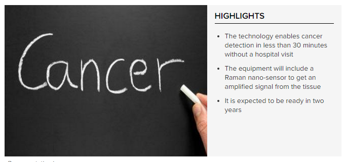 COIMBATORE: Four years ago, Shantikumar V Nair and Manzoor Koyakutty of Amrita University's Kochi-based Centre for Nanomedicine were using lasers to detect food contaminants using Raman spectroscopy. As the contaminants threw up distinct patterns, Nair wondered why not bounce lasers off human tissue to detect cancercells?
The result is a technology that enables cancer detection in less than 30 minutes without a hospital visit. The team is now working on a hand-held instrument to detect oral cancer. It is expected to be ready in two years. "We used the method on samples of oral cancer, normal tissues and pre-cancerous tissues. The first set of positive results came two years ago," said Shantikumar V Nair, one of the three inventors of the technology. The department of biotechnology has provided the team Rs 60 lakh to design the gadget, expected to cost around Rs 10 lakh. The cancer detection technology uses a laser with a nano substrate to detect pre-cancerous and cancerous cells. Use of laser to read the nature of cells is not new, but the signals that they produced were found to be weak and difficult to analyse. The nano substrate (a nano material) placed on the tissue solved the problem. "The nano substrate amplifies the signals and helps us analyse the results. For each type of tissue — normal, pre-cancerous and cancerous, there is a distinct Raman spectrum emitted by the laser," said Nair. "Raman signal helps identify organic molecules in tissues. Since these molecules vary depending on the condition of the cells, signals coming from cancer tissue with those coming from normal tissue will be distinct," said Nair. The equipment will include a Raman nano-sensor to get an amplified signal from the tissue. The signals can be analysed at a central facility, and the result will be available within 30 minutes. While this is a preliminary test to detect cancer, scientists say gold-standard tests will have to confirm it. Nair, who is also the dean of research at Amrita University, said the new gadget will enable community level large-scale screening without having to take tissue biopsies. "In principle, it can diagnose any cancer from which Raman spectra can be obtained... We have paid more attention to oral cancer because of the possibility of screening for these cancers early and also because oral cancer is on the rise and is curable if detected early," Nair said. Amrita Nano Centre Scientists Win International Patents for Novel Invention on Cancer-Nanomedicine8/16/2016  Amrita Nano Centre Scientists Win International Patents for Novel Invention on Cancer-NanomedicineAmrita Centre for Nanoscience and Molecular Medicine, Kochi scientists lead by Prof. Manzoor Koyakutty and Prof. Shantikumar Nair (Centre Director) received two international patents from USA, Europe, Japan and China for a path-breaking invention in cancer-nanomedicine for treating drug-resistant cancers. Nanomedicines are tiny nanoparticles (one billionth of a meter) loaded with drug molecules to efficiently treat various types of diseases. This new nanomedicine invented by Amrita team is specifically designed to identify resistant cancer cells and deliver more than one drug's molecules simultaneously to stop multiple deregulated cancer mechanisms. “For the last 5 years, in collaboration with Dr. K. Pavithran, Dr. Neeraj Sidharthan, and Dr. Ranvir Prabhu, at the Medical-Oncology department of Amrita Institute of Medical Sciences (AIMS), we were investigating key molecular mechanisms responsible for the chemo-resistance of various types of cancers,” said Dr. Manzoor K., who led the nanomedicine research with Dr. Nair. “Based on our understanding, we have designed a novel core-shell nanosystem using human proteins and biodegradable polymers to target cancer cells while causing little harm to other healthy organs,” he further explained. “This patent is the first of its kind in the field of ‘Cancer-nanomedicine’ patents won by Indian scientists competing at the international level and Amrita is proud to make such a significant lead in in the emerging area of cancer-nanotechnology,” said Dr. Shantikumar Nair, Director of Amrita Center for Nanosciences and Molecular Medicine. “Currently, advanced chemodrugs are unaffordable for the common public because most of the inventions are made in USA or Europe. In contrast, our invention is made completely in India and hence this can be made available to the common man at an affordable cost,” added Dr. Nair. Presently, Amrita is conducting safety trials in animal models which are mandatory to start human clinical trials. These studies are expected to be completed within next 3-4 years. PhD researchers Dr. Parvathy Chandran, Dr. Archana Ratnakumari and Giridharan Malarvizhi participated in this invention. Other team members- Dr. Anusha Ashokan, Dr. Girish, Dr. Ranjith, Dr. Vijay Harish, Dr. Jeena, Lakshmi G. Kumar, Manju C. Abraham, Jyotsna, are actively involved in various translational aspects of this innovation. The project was sponsored by the Department of Biotechnology, Government of India. US PATENT: Title: Core-Shell Particle Formulation for Delivering Multiple Therapeutic Agents Inventors: Manzoor Koyakutty, Parvathy Chandran , Giridharan L M, Archana Retnakumary and Shanti Nair, Patent No: USPTO 9,402,918. 2016 EUROPEAN PATENT: No. 09788193.2-1453 JAPAN PATENT: No. JP5662431, China: Zl2009 8.0160922.6 Title: Targeted Nanophotomedicine for the photodynamic therapy of Cancer Inventors: Manzoor Koyakutty, Shantikumar Nair et al. https://www.amrita.edu/news/amrita-nano-centre-scientists-win-international-patents-novel-invention-cancer-nanomedicine
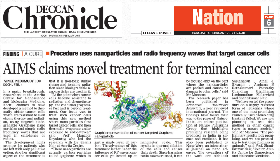
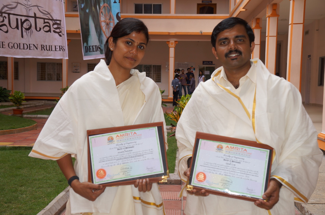
Congratulations to Dr. Manzoor and team for being awarded two international patents for their work!!4/6/2016 Amrita Centre for Nanoscience and Molecular Medicine, Kochi scientists lead by Prof. Manzoor Koyakutty and Prof. Shantikumar Nair (Centre Director) received two international patents from USA, Europe, Japan and China for a path-breaking invention on cancer-nanomedicine for treating drug-resistant cancers. Nanomedicines are tiny nanoparticles (one billionth of a meter) loaded with drug molecules to efficiently treat various types of diseases. This new nanomedicine invented by Amrita team is specifically designed to identify resistant cancer cells and deliver more than one drug molecules simultaneously to stop multiple deregulated cancer mechanisms. ‘From last 05 years, in collaboration with Dr, K Pavithran, Dr Neeraj Sidharthan, and Dr Ranvir Prabhu, at medical-oncology department of AIMS, we were investigating key molecular mechanisms responsible for the chemo-resistance of various types cancers, said Dr Manzoor K, who led the nanomedicine research with Dr Nair. Based on our understanding, we have designed a novel core-shell nanosystem using human proteins and biodegradable polymers to target cancer cells while causing little harm to other healthy organs. ‘This is the first of its kind ‘Cance-nanomedicine’ patents won by Indian scientists by competing at international level and Amrita is proud to make such a significant lead in in the emerging area of cancer-nanotechnology, said Dr Shanti Nair, Director of Amrita Nanocentre. ‘Currently, advanced chemodrugs are unaffordable for common public because most of the inventions are made in USA or Europe. In contrast, our invention is made completely in India and hence this can be made available to common man at affordable cost, said Dr Nair. Presently Amrita is conducting safety trials in animal models which are mandatory to start human clinical trials. These studies are expected to be completed within next 03-04 years. PhD researchers Dr Parvathy Chandran, Dr Archana Ratnakumari and Giridharan Malarvizhi participated in this invention. Other team members Dr Anusha Ashokan, Dr Girish, Dr Ranjith, Dr Vijay Harish, Dr Jeena, Lakshmi G Kumar, Manju C Abraham, Jyotsna are actively involved in various translational aspects of this innovation. The project was sponsored by Department of Biotechnology, Govt of India. Students Anjana, Ida received their MTech and Anusha, Girish received PhD degrees during 12th convocation of the Amrita university. A new article photo feature with the our Research Scientist Dr Anusha. 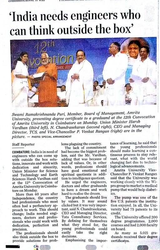  doi:10.1038/nindia.2015.13 Published online 29 January 2015 How does one stop the runaway growth of cancer cells that defy anticancer drugs and radiation therapy? An Indian research team has found a way to stifle the growth of drug- and radiation-resistant cancer cells. They have developed graphene nanoparticles that can convert low radiofrequency powers to heat which annihilates such chronic myeloid leukaemia cells1.
“This combination of graphene nanoparticles and radiofrequency power is a promising cancer therapy as it offers minimally invasive treatment without the need of other therapeutic aids,” says first author Abhilash Sasidharan from Amrita Centre for Nanoscience and Molecular Medicine (ACNSMM), Kerala and now a post-doctoral fellow at the US-based Kansas State University. The therapy could potentially be used to selectively kill other types of drug-resistant cancer cells, he told Nature India. Due to rise in drug- and radiation-resistant cancer cases, scientists have been trying to develop alternative anticancer therapies. They had synthesized carbon nanotubes and gold nanoparticles able to convert radio waves and near-infrared light to heat that killed cancer cells. But these nanoparticles generated heat that only killed malignant tumours smaller than 5 cm. For larger tumours that could spread to multiple organs, Sasidharan along with ACNSMM director Shantikumar Nair and senior scientist Manzoor Koyakutty set out to probe the heat-generating potential of surface-modified graphene nanoparticles by exposing them to low radiofrequency powers. The team found that when exposed to low radiofrequency powers (10-50 watts) for 5 minutes, graphene showed higher thermal responses than those of single-walled carbon nanotubes and gold nanoparticles. “These higher thermal responses of graphene may be attributed to high electron density on the graphene surface which, in turn, leads to high current densities, resulting in enhanced heat production,” Koyakutty explains. The scientists then investigated whether these graphene nanoparticles could kill drug- and radiation-resistant cancer cells by converting low radiofrequency powers to heat. They attached the graphene nanoparticles with transferrin, an iron-transport protein. This protein binds to receptors which are highly expressed on the surface of drug-resistant cancer cells. Next, they exposed graphene-treated drug- and radiation-resistant cancer cells of chronic myeloid leukaemia to radiofrequency power of 50 watts for 5 minutes. Sophisticated imaging techniques revealed that graphene treatment followed by exposure to radiofrequency power triggered controlled death of the cancer cells. In a separate animal study, the team had earlier shown that similar graphene nanoparticles are biodegradable and not-toxic to living cells2. “These results indicate that the combination of graphene nanoparticles and radiofrequency power could lead to effective cancer therapy for combating drug- and radiation-resistant cancers,” Koyakutty says. Asifkhan Shanavas from the Institute of Nano Science and Technology, Punjab says the therapy precisely targets cancer cells of chronic myeloid leukaemia and also enhances the efficacy of radiation therapy, an existing cancer treatment. The graphene nanoparticles’ ability to kill tough cancer cells is encouraging since treating cancers with small drug molecules always results in the emergence of new drug-resistant mechanisms, he adds. References 1. Sashidharan, A. et al. Radiofrequency ablation of drug-resistant cancer cells using molecularly targeted carboxyl-functionalized biodegradable graphene. Adv. Health. Mat.(2015) doi:10.1002/adhm.201400670 2. Girish, C. M. et al. Confocal raman imaging study showing macrophage mediated biodegradation of graphene in vivo. Adv. Health. Mat. 2, 1489-1500 (2013) This is an important article for all those interested to know more about Cancer detection by testing blood / tissue samples
|
AuthorDr Manzoor et al., Archives
October 2017
Categories
|
 RSS Feed
